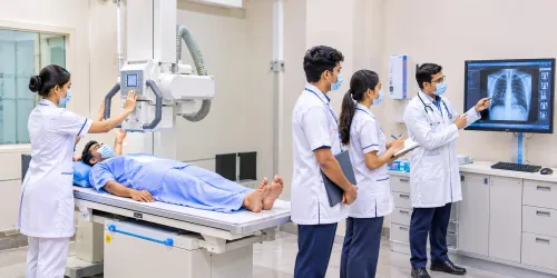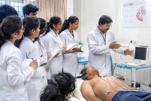


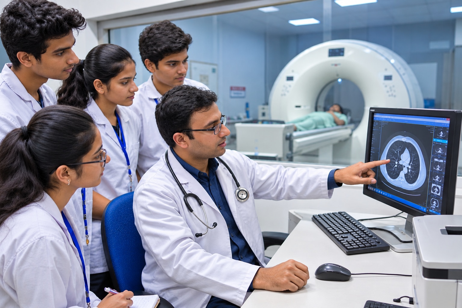
A DRIT syllabus can look comprehensive while offering very different levels of practical exposure, modality training and workplace readiness. By comparing its subjects, clinical hours, equipment access and assessment methods, you can judge which technical skills support entry-level imaging work and which require further supervised training.
Key takeaways
- Choose programs with supervised practice across X-ray, CT, MRI and ultrasound.
- Confirm students perform patient positioning, exposure selection and image quality checks.
- Ask for radiation-safety training, dosimetry procedures and quality-control records.
- Compare clinical placements, equipment access and career pathways before enrolling.
What does a term-by-term DRIT curriculum actually teach?
A conventional DRIT curriculum moves from scientific foundations to supervised imaging practice. Each term adds a skill needed to produce safe, diagnostically useful images and work effectively with patients and clinical teams.
- Term 1: Anatomy and physiology identifies bones, organs, body systems and anatomical landmarks. Patient care covers identification, communication, consent checks, mobility support and basic infection control.
- Term 2: Radiation physics explains X-ray production, exposure factors, beam characteristics and how distance, shielding and collimation affect dose. Radiation protection connects these principles to safe daily practice.
- Term 3: Radiographic positioning teaches projections, body alignment, anatomical markers, immobilisation and motion control. Students learn how poor positioning creates repeat images and unnecessary patient exposure.
- Term 4: Image processing and quality assessment cover exposure indicators, contrast, sharpness, artefacts and rejection analysis. Equipment operation includes the console, detector, grid, tube movement and portable X-ray workflow.
- Term 5: Clinical postings bring these skills together during supervised examinations, including patient preparation, positioning, exposure selection, image evaluation and documentation. Emergency, bedside and infection-control procedures expose students to real workflow pressure.
- Term 6: CT, MRI, ultrasound, fluoroscopy, mammography, dental imaging and contrast examinations may appear as modules or introductory rotations. Treat observation as exposure, not proof of independent competence.
By the end, employable competence means completing assigned examinations safely under the permitted level of supervision. A diploma does not authorise diagnosis or radiology reporting, and advanced modality roles require documented experience or further training.
Which procedures should a DRIT student perform before working independently?
A student should work independently only after completing the full examination workflow safely, not after memorising positioning charts. The required sign-off covers patient identification, consent checks, patient care, positioning, exposure selection, image evaluation, documentation and escalation of problems.
| Stage | What the student must demonstrate | Appropriate responsibility |
|---|---|---|
| Classroom theory | Explain anatomy, physiology, radiographic physics, radiation protection, exposure factors, image quality and ethics | No patient examination |
| Simulation | Identify the patient and examination, practise positioning, marker placement, collimation, infection control and equipment operation | Perform on a model or simulator with instructor correction |
| Supervised clinical posting | Manage real patients, including children, anxious patients and mobility-limited patients; operate the assigned system, review images and report faults | Perform examinations only with a qualified technologist observing and signing off |
| Independent work | Complete approved examinations, verify worklists and DICOM details, evaluate diagnostic adequacy, document the procedure and escalate repeats, incidents or equipment faults | Work alone only within the signed competency list |
Before sign-off, the student should repeatedly complete:
- Confirm two patient identifiers, the requested examination, relevant history and pregnancy-related information under local policy.
- Explain the procedure, complete consent checks where required, protect privacy and use safe transfers or positioning supports.
- Select exposure factors, collimate and control motion without creating avoidable repeat exposures.
- Inspect image evaluation criteria: anatomy included, positioning, marker placement, exposure, artefacts and diagnostic usability.
- Follow cleaning and infection-control procedures, then record the examination and report discrepancies.
A posting that offers observation without documented supervised examinations does not prove readiness. CT, MRI, ultrasound, mammography, fluoroscopy and dental imaging require separate competency sign-offs; classroom introduction alone does not authorise independent operation.
Which imaging modalities, equipment and digital systems belong in the training?
Plain radiography should be the core hands-on competency: students need to position patients, select exposure factors, operate fixed and portable units, assess images, and repeat only for a documented reason. Training must cover both CR and DR systems, image receptors, processing errors, artefacts, and safe equipment shutdown.
| Modality | Skills and equipment to learn | Expected training level |
|---|---|---|
| Plain radiography | Fixed and mobile X-ray units, CR and DR systems, grids, exposure controls and positioning | Full supervised competence |
| CT | Gantry, console, protocols, dose indicators, patient preparation and contrast workflow | Supervised practice; independent scanning requires documented authorization |
| MRI | Magnet-room zones, screening, implants, coils, sequences and emergency quench procedures | Introductory or closely supervised exposure |
| Ultrasound | Transducers, machine controls, Doppler, image labeling and patient preparation | Introductory scanning; reporting is not a technologist function |
| Mammography | Dedicated unit, compression, positioning, exposure and privacy requirements | Introductory exposure unless a placement provides specialist training |
| Fluoroscopy | C-arm or remote fluoroscopy equipment, timed exposure, contrast assistance and sterile workflow | Supervised assistance |
| Dental imaging | Intraoral sensors, panoramic and cephalometric units, positioning and infection control | Introductory or supervised practice |
Digital workflow belongs in every modality. Students should use modality worklists, verify two patient identifiers, transfer studies through DICOM, and understand PACS and RIS. A technically good image attached to the wrong patient is still a serious failure.
Clinical postings must show actual examinations, not only demonstrations. MRI, CT, ultrasound imaging courses, mammography, fluoroscopy and dental imaging may introduce equipment and safety without granting independent competence.
How do radiation safety and quality control shape daily imaging work?
A safe imaging department applies the ALARA principle while producing a diagnostic image on the first attempt. Radiation protection means controlling exposure through time, distance and shielding, then reducing repeats with correct positioning, exposure factors and collimation.
- Spend less time near the source, maximise distance during exposure and use structural barriers or protective aprons when appropriate.
- Collimate to the anatomy requested; an open field increases dose and scatters radiation into the room.
- Wear a personal dosimeter in the assigned position and review its readings. It records occupational exposure but does not protect you or replace shielding and correct technique.
- Ask pregnancy-related questions under the facility’s policy before examinations involving ionising radiation, and escalate uncertainty before exposing the patient.
- For contrast studies, confirm patient identity, examination indication, previous contrast reactions, relevant kidney-function requirements and intravenous access. Keep emergency equipment and trained assistance available according to department protocol.
- Clean contact surfaces, use hand hygiene and select infection-control precautions for every patient, especially during portable, emergency and isolation-room imaging.
The Radiation Safety Officer manages the protection programme, staff training, dose monitoring, incident investigation and compliance audits. Students should know how to report an unsafe exposure rather than conceal it.
Quality-control checks protect both patients and diagnostic accuracy. Staff inspect equipment condition, detector uniformity, exposure indicators, image artefacts, calibration records and reject patterns; scheduled tests also verify generator and tube performance. A failed check can create repeated images or hide a fault, so the equipment is removed from use and reported according to procedure.
How can you compare DRIT programs and connect training to career options?
A low fee or long subject list does not prove a strong DRIT program. Compare documents that show where training happens, who assesses you, which body awards the qualification, and whether the relevant allied-health regulator accepts it.
Ask the institute for:
- The published syllabus, clinical-hour requirements, attendance rules and examination scheme.
- A hospital attachment letter naming the hospital, department, placement dates and supervised examinations.
- A student logbook showing procedures, case numbers, supervisor sign-offs and incident reporting.
- The awarding authority, accreditation or affiliation document, and written evidence of regulator recognition.
- A modality list stating whether students observe, simulate or perform work on X-ray, CT, MRI, ultrasound, mammography, fluoroscopy and dental systems.
- Evidence that training includes PACS and DICOM workflows, RIS worklists, patient identification and image transfer.
| Criterion | Strong evidence | Warning sign |
|---|---|---|
| Clinical exposure | Named hospital, logged examinations and direct supervision | “Practical training” with no hours, cases or placement site |
| Assessment | Practical examinations, image-evaluation tasks and documented competency sign-offs | Marks based only on written theory |
| Awarding authority | Verifiable certificate issuer and regulator acceptance | Unclear affiliation or registration eligibility |
| Modality access | Timetabled access with supervised cases, not just demonstrations | Equipment listed but no student access |
This evidence links directly to work. A sound foundation can support entry-level general radiography, portable and emergency imaging, patient preparation, image processing, or supervised assistance in CT and MRI; it does not authorise diagnosis or radiology reporting.
Later career progression in radiology may lead to CT, MRI, mammography, quality assurance or applications work after additional training and supervised experience. KP PARAMEDICAL INSTITUTE PCMC is worth comparing against this checklist rather than choosing from its course title alone.
Related product
 | Best DRIT Course The DRIT (Diploma in Radiology & Imaging Technology) at KP Paramedical Institute PCMC is a career-oriented paramedical program designed to prepare... View product → |
Frequently asked questions
What does a term-by-term DRIT curriculum teach?
It progresses from anatomy, physiology and radiation science to patient care, imaging procedures, image evaluation and clinical placement.
Which procedures should DRIT students perform before working independently?
Students should complete supervised practice in patient identification, positioning, exposure selection, equipment operation, image evaluation and infection control.
Which imaging modalities and digital systems belong in DRIT training?
Training should cover X-ray and digital radiography, with exposure to CT, MRI, ultrasound, PACS, RIS and DICOM workflows.
How do radiation safety and quality control shape imaging work?
Daily work includes justification, collimation, protective measures, dose monitoring, equipment checks, reject analysis and documented quality control.