


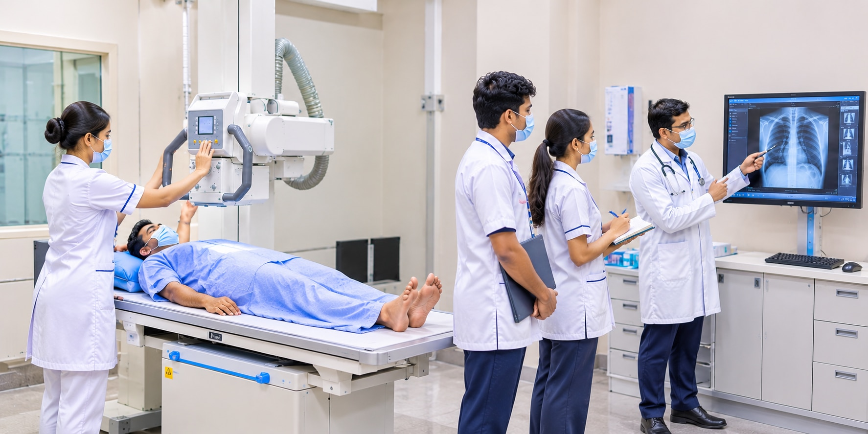
An X-ray is more than a quick image: the tube, detector, positioning, exposure settings and image processing all affect what a clinician can see. By the end, you will know when radiography is useful, when another test is better, how an examination is performed, and which safety practices protect patients without delaying necessary care.
Key takeaways
- Bone absorbs more X-rays than air or soft tissue, creating visible image contrast.
- Use X-rays for many bone and chest assessments, but choose CT, MRI, or ultrasound when detail demands it.
- Tell staff if you are pregnant or might be pregnant before the examination.
- Modern detectors, exposure controls, and trained interpretation support safer, more useful imaging.
How an X-ray becomes a diagnostic image
An X-ray image is a map of how much radiation reaches a detector. The X-ray tube sends X-ray photons through the body; bone absorbs more photons than air or soft tissue, so different tissues create contrasting areas. The detector converts that pattern into an electronic image, which software adjusts for brightness and contrast.
Patient positioning determines whether anatomy is projected clearly or hidden by overlap. Rotation can make the heart appear larger, obscure a fracture, or mimic abnormal alignment. Motion blurs edges, while incorrect centring or collimation can exclude the area that needs assessment.
A useful radiograph needs:
- The correct body part and view for the clinical question.
- Sharp detail without motion blur.
- Adequate exposure and contrast.
- Accurate markers, patient identity and side designation.
Digital systems can make an overexposed image look acceptable, so a bright picture does not prove that the dose was appropriate. Exposure indicators, technique charts and avoiding unnecessary repeats support quality control. Portable examinations help image patients who cannot be moved, but limited positioning can reduce diagnostic confidence and require correlation or repeat imaging.
A radiograph supports the history and examination; a technically good image still needs clinical interpretation.
What X-rays show—and when another test is better
Plain radiography can identify fractures, dislocations, bowel obstruction, dental abnormalities and many lung findings. A chest X-ray can support diagnosis of pneumonia, tuberculosis or pneumothorax, but a normal image does not exclude an early infection, small lesion or occult fracture.
| Test | Best at showing | When clinicians choose it |
|---|---|---|
| Plain radiography | Bone alignment, lung fields, teeth and bowel gas patterns | First-line assessment for many injuries and chest symptoms |
| Fluoroscopy | Moving anatomy and contrast flow | Swallowing studies, gastrointestinal examinations and some procedures |
| Mammography | Fine breast densities and calcifications | Breast screening or evaluation of a lump |
| CT | Cross-sectional bone, lung, abdomen and bleeding | Complex fractures or emergencies when radiographs are insufficient |
| MRI | Ligaments, tendons, brain, spinal cord and other soft tissue | Suspected soft-tissue injury or disease; it uses no ionizing radiation |
| Ultrasound | Fluid, gallbladder, vessels, pregnancy and superficial tissue | Real-time imaging without ionizing radiation |
| Laboratory testing | Blood, urine or tissue abnormalities | Infection, anemia, organ dysfunction or diseases that imaging cannot confirm |
CT also uses X-rays but delivers a higher dose than a plain examination; an adult two-view chest X-ray is about 0.1 mSv, while a chest CT is about 6.1 mSv, with actual dose varying by protocol.
Clinicians select the least burdensome test that answers the clinical question, then escalate when symptoms and the first result disagree. Fluoroscopy and some angiographic studies add iodinated or barium contrast, requiring checks for allergies, kidney function, aspiration and perforation risks.
What happens during an X-ray examination
A clinician’s referral states the body part and clinical question. Before imaging, the radiographer confirms your name, date of birth, examination, and relevant history, including pregnancy possibility, previous images, and your ability to stand, sit, or lie still.
- Preparation: Remove jewellery, clothing fasteners, or other objects over the area. Follow instructions about breathing, keeping still, or holding a position.
- Positioning: The radiographer places you and the body part against the detector, using markers and supports when needed. Correct alignment prevents rotation and hidden anatomy.
- Exposure: You remain still while the X-ray technician selects exposure factors such as tube voltage, current, and exposure time, then makes the image in a fraction of a second. You are not radioactive afterward.
- Image review: The radiographer checks coverage, positioning, motion, markers, and image quality before you leave. A technically limited image can hide disease or require a repeat; portable examinations can face extra positioning limits when moving you is unsafe.
- Reporting: A radiologist reviews the images alongside the referral and any prior studies, then sends a report to the clinician. The report supports diagnosis but does not replace your symptoms, examination, or other tests.
Most patients return to normal activities immediately. Ask when and how your referring clinician will explain the result, because the radiographer usually cannot provide the final diagnosis during the examination.
How radiation safety protects patients without blocking needed care
A medically justified X-ray balances a real diagnostic benefit against a small radiation risk, rather than avoiding exposure at any cost. ALARA—“as low as reasonably achievable”—means using the lowest dose that produces an image good enough to answer the clinical question.
- Justification: confirm that the examination is needed and that another test will not answer the question better. This prevents unnecessary imaging.
- Time: keep exposure time and the time spent near an active X-ray beam as short as practical.
- Distance: staff step away or use remote controls during exposure; doubling distance from the source sharply reduces their exposure.
- Shielding: protective barriers and, when appropriate, shields reduce exposure to staff and nearby people. Shielding never compensates for poor technique.
- Collimation: restrict the beam to the body area being examined. A smaller field reduces dose and improves image contrast.
- Repeat-image reduction: position carefully, prevent motion, check exposure settings and review the image before the patient leaves. A technically poor image can hide disease and force another exposure.
Paediatric imaging uses child-sized exposure settings because children are more radiation-sensitive and have longer lifetimes for effects to develop. Never apply adult settings automatically.
Pregnancy screening identifies a known or possible pregnancy before imaging. Pregnancy does not automatically cancel a necessary X-ray; the team can adjust justification, collimation, positioning and protocol, with particular care for abdominal or pelvic studies. Tell the healthcare team before the examination.
Why modern X-ray services depend on technology and trained judgment
A technically acceptable image is not automatically a diagnostically useful one. Digital radiography gives rapid images and supports exposure monitoring, but its wide exposure range can hide overexposure; technique charts, exposure indicators and repeat analysis remain essential.
Computed radiography uses a reusable imaging plate, adding a readout step that can delay workflow and introduce plate or reader errors.
| Method | What it adds | Quality decision |
|---|---|---|
| Fluoroscopy | Live X-ray guidance for swallowing studies, contrast examinations and procedures | Balance motion detail against cumulative exposure |
| Mammography | High-detail breast imaging using dedicated views and compression | Control positioning, compression and artifacts |
| Portable imaging | Images patients who cannot safely reach the radiography room | Accept limited positioning or request a repeat when it changes interpretation |
Portable equipment is valuable in emergency and intensive-care settings, yet bed angle, tubes, rotation and weaker positioning can obscure disease. Radiology review checks exposure, anatomy, artifacts and prior images before interpretation; the report still needs symptoms and examination findings to give a shadow clinical meaning.
Training must therefore cover equipment operation, positioning, radiation control, image critique and communication—not button pressing alone. When comparing courses, check whether practical learning includes rejecting a blurred or rotated image for a clear reason; KP PARAMEDICAL INSTITUTE PCMC is relevant to that decision because imaging training should connect technical faults with patient-care consequences.
Related products
 | Best X-Ray Technician Course The Diploma in X-Ray Technology at KP Paramedical Institute PCMC is a career-oriented paramedical program designed to prepare students for careers in... View product → |
 | Best DRIT Course The DRIT (Diploma in Radiology & Imaging Technology) at KP Paramedical Institute PCMC is a career-oriented paramedical program designed to prepare... View product → |
Frequently asked questions
How does an X-ray become a diagnostic image?
An X-ray tube sends photons through the body, and a detector records how many reach it. Bone absorbs more photons than air or soft tissue, creating contrast that software adjusts for brightness and detail.
What do X-rays show, and when is another test better?
X-rays show bones, chest structures, and differences in tissue density. CT provides more detailed cross-sectional views, MRI shows soft tissues without ionising radiation, and ultrasound helps assess moving or fluid-filled structures.
What happens during an X-ray examination?
You remove objects that could block the image, follow positioning instructions, and remain still while the technologist takes one or more exposures. The examination is usually brief and painless.
How does radiation safety protect patients during an X-ray?
Staff use the lowest exposure that produces a diagnostic image, limit the scanned area, check pregnancy status when relevant, and avoid repeat images through careful positioning and technique.